The Ultimate Guide to Pet Dental Care

Pet dental care is one of the most overlooked aspects of pet care. According to the American Veterinary Dental Society, over 80% of dogs and 70% of cats show signs of periodontal (gum) disease by age three.

Because pets rarely complain about dental pain, oral disease often progresses unnoticed until it causes severe discomfort, tooth loss, or systemic health issues affecting the heart, liver, and kidneys.

Why Pet Dental Care Matters

When a pet eats, bacteria combine with food particles and saliva to form a soft film called plaque. If plaque isn’t brushed away within 24–48 hours, it mineralizes with salivary calcium to form hard calculus (tartar).

       Plaque Formation          Tartar Mineralization         Periodontal Disease
  [Bacteria + Food Residue]  ──>  [Calcified Hard Crust]  ──>  [Bone & Attachment Loss]
       (Reversible)                   (Requires Scaling)             (Irreversible)

Tartar creates a rough surface where more bacteria attach, migrating under the gumline. Left untreated, the body’s inflammatory response leads to:

  • Gingivitis: Red, swollen gums that bleed easily (reversible).

  • Periodontitis: Destruction of the periodontal ligament and supporting alveolar bone, leading to tooth mobility and eventual loss (irreversible).

  • Systemic Organ Damage: Oral bacteria can enter the bloodstream through inflamed gums, spreading to vital organs like the heart valves, liver, and kidneys.

Understanding the Stages of Dental Disease

Veterinarians classify dental disease into four distinct grades:

Stage Description Reversible? Treatment Required
Grade 1 (Gingivitis) Mild gum redness; soft plaque build-up; no attachment loss. Yes Home care & professional polish
Grade 2 (Early Periodontitis) Inflamed gums; early pocket formation; less than 25% bone loss. No (Controlled) Professional dental scaling under anesthesia
Grade 3 (Moderate Periodontitis) Thick tartar crust; swollen/bleeding gums; 25%–50% bone loss. No Scaling, root planing, or extraction
Grade 4 (Advanced Periodontitis) Severe tissue destruction; pus around gums; greater than 50% bone loss; loose teeth. No Surgical tooth extractions required

Pet Dental Care

Daily Prevention: How to Brush Your Pet’s Teeth

Daily mechanical brushing remains the gold standard for home dental care.

CRITICAL WARNING: Never use human toothpaste on pets. Human toothpastes often contain xylitol (a sweetener highly toxic to dogs causing fatal hypoglycemia and liver failure) or high levels of fluoride, which causes severe gastric distress when swallowed. Always use enzymatically active, pet-safe toothpaste (e.g., poultry or malt flavor).

Step-by-Step Brushing Protocol

1. Desensitization & Flavor Association: Days 1–3.

Let your pet lick a small dab of pet-safe enzymatic toothpaste directly off your finger. Reward them immediately with praise to build a positive association.

2 . Finger Gum Rubs: Days 4–7.

Apply toothpaste to your finger or a soft silicone finger-brush. Gently lift their upper lip and rub the outer surfaces of the upper canine teeth and molars in a circular motion for 10–15 seconds.

3.Introduce a Pet Toothbrush:  Week 2 Onward.

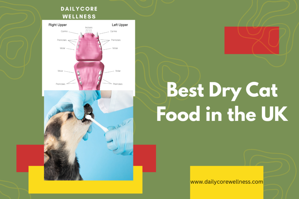
Transition to an angled pet toothbrush held at a 45-degree angle to the gumline. Focus primary effort on the outer surfaces of the upper back teeth (where tartar accumulates fastest due to salivary gland openings).

Complementary At-Home Dental Products

If your pet refuses toothbrushing, combining passive dental aids can still slow down plaque accumulation:

  1. VOHC-Approved Dental Chews: Look for the Veterinary Oral Health Council (VOHC) seal of acceptance on packaging. VOHC-certified chews (like Greenies or Oravet) are scientifically proven to reduce plaque or tartar by specific margins.

  2. Water Additives & Enzymatic Sprays: Added to fresh drinking water daily, these additives contain chlorhexidine or natural enzymes that reduce oral bacterial loads.

  3. Prescription Dental Diets: Specialized kibbles (such as Hill’s t/d or Royal Canin Dental) feature oversized, fiber-aligned kibbles that scrape the tooth surface clean like a squeegee before shattering.


Advanced Pet Dental Care: Recognizing Pain and Understanding Professional Cleanings

While home brushing and dental chews slow down plaque accumulation, every pet eventually requires professional veterinary care. Because pets are master survivalists who instinctively mask chronic pain to avoid showing vulnerability, recognizing oral disease requires looking beyond simple bad breath.

Recognizing Hidden Signs of Dental Pain

Dogs and cats rarely stop eating entirely due to a toothache; instead, they alter how they eat and behave. Look out for these subtle red flags:

  • Dropping Food or “Chomping”: Picking up kibble, dropping it back out of the mouth, or tilting their head sideways to chew exclusively on one side.

  • Behavioral Shifts: Sudden head shyness, snapping or pulling away when touched near the muzzle, or uncharacteristic lethargy.

  • Pawing at the Face: Frequently rubbing their mouth against carpets, furniture, or using their paws to scratch at the jaw.

  • Excessive Salivation & Halitosis: While mild dog breath is common, persistent, rotting odors indicate active bacterial infection, periodontal pockets, or dying tooth pulp.

See also  The Complete Guide on Pets4Homes UK

The Truth About Professional Veterinary Dental Cleanings

Many pet owners encounter advertisements for “anesthesia-free teeth cleaning” at grooming salons. Veterinary dental organizations universally condemn this practice for two critical reasons:

  1. Invisible Disease: Over 60% of a dog or cat’s tooth structure resides below the gumline. An awake pet cannot tolerate subgingival scaling, root planing, or diagnostic dental X-rays.

  2. Trauma and Stress: Scraping teeth on an awake, terrified animal removes only the cosmetic tartar on the crown while leaving painful, bacteria-laden subgingival pockets untouched, causing immense psychological stress.

A safe, comprehensive veterinary dental cleaning requires general anesthesia with endotracheal intubation (to protect the airway from water and debris) and advanced multimodality pain management.

Step-by-Step Anatomy of a Professional Vet Dental Procedure

1. Step 1: Pre-Anesthetic Bloodwork & Charting: Comprehensive Assessment.

Your vet runs blood panels to verify organ function before anesthesia, then records missing, fractured, or mobile teeth on a specialized dental chart.

2.Step 2: Full-Mouth Dental Radiography: Intraoral Imaging.

X-rays reveal hidden problems beneath the bone—such as tooth root abscesses, bone loss, or resorptive lesions (common in cats)—that are invisible to the naked eye.

3.Step 3: Crown & Subgingival Cleaning: Ultrasonic Scaling.

An ultrasonic scaler vibrates at high frequency to shatter calculus above and below the gumline, followed by fine hand-scaling in deep periodontal pockets.

4.Step 4: Tooth Polishing:  Polishing & Fluoride.

Scaling leaves microscopic scratches on the enamel. Medical-grade polishing smooths the surface to delay future plaque re-attachment.

5.Step 5: Extractions & Advanced Treatment: Surgical Intervention.

If teeth are unsalvageable due to advanced bone loss or feline tooth resorption, localized nerve blocks are administered, and diseased teeth are surgically extracted.

Breed-Specific Dental Challenges and Feline Tooth Resorption

Understanding pet dental health requires looking at how genetics and species-specific pathology affect oral care. Certain dog breeds face severe structural disadvantages due to skull shape, while cats suffer from a unique, painful dental condition that differs entirely from standard periodontal disease.

1. Brachycephalic Dental Overcrowding (Pugs, Bulldogs, & Persians)

Brachycephalic pets—animals bred to have shortened, flat facial structures—possess the same number of teeth as their long-snouted counterparts (42 permanent teeth in dogs, 30 in cats). However, these teeth are forced to fit into a significantly compressed jawbone.

The Structural Problem

When 42 full-sized adult teeth attempt to erupt within a shortened maxilla (upper jaw), several anatomical complications arise:

  • Rotated & Malaligned Teeth: Premolars often erupt sideways or at 90-degree angles to fit into the dental arch.

  • Elimination of Interdental Spaces: Healthy teeth have gaps between them, allowing saliva to wash away food debris. Overcrowded teeth touch side-by-side, creating severe plaque-trapping crevices.

  • Unerupted or Impacted Teeth: Teeth can remain trapped under the gumline, leading to the formation of painful dentigerous cysts that destroy surrounding jawbone tissue over time.

Preventative Strategy: Brachycephalic breeds require daily brushing starting in puppyhood/kittenhood and often benefit from early veterinary evaluations (around 6–9 months of age) to extract retained deciduous (baby) teeth or severely rotated premolars before localized bone destruction occurs.

2. Feline Tooth Resorption (FORL)

Unlike periodontal disease, which is driven by external bacterial plaque, Tooth Resorption (TR) is an internal, progressive condition where a cat’s own body begins breaking down and absorbing the structural tissues of its teeth. It affects 30% to 60% of adult cats over the age of five.

How Resorption Works

Specialized cells called odontoclasts (which normally dissolve baby tooth roots during kittenhood) inexplicably reactivate in adult cats. They erode the protective enamel and underlying dentin near the gumline:

  1. Stage 1–2 (Mild to Moderate): Eosinophilic pink tissue (granulation tissue) grows over the crown as the enamel wears away, exposing the sensitive innervated dentin underneath.

  2. Stage 3–4 (Advanced): The root structure degrades significantly, often fusing directly with the surrounding alveolar bone (ankylosis) or leaving painful, exposed nerve pulp inside the mouth.

  3. Stage 5 (End-Stage): The entire crown shatters, leaving only remnant root fragments buried under inflamed gum tissue.

See also  Complete Guide to The Best Wet Cat Food in the UK

Clinical Management

Tooth resorption cannot be prevented or reversed by toothbrushing alone because its root etiology remains largely idiopathic (unknown). Because the condition is extremely painful, treatment requires veterinary intervention under anesthesia:

  • Type 1 Resorption (Normal Root Structures): Complete surgical extraction of the tooth and its intact roots.

  • Type 2 Resorption (Roots Replaced by Bone): Crown amputation, where the exposed crown is removed, and the degrading roots are allowed to safely absorb into the jawbone under closed gum flaps.

3. Post-Operative Care After Pet Tooth Extractions

When a pet undergoes dental extractions, proper home care during the 10–14-day healing window determines whether surgical sites close cleanly or develop painful infections.

1.  Phase 1: Pain Management & Ultra-Soft Food: Days 1–3 Post-Op.

Administer all prescribed pain medications and antibiotics exactly as directed by your vet. Feed strictly soft food (canned food or kibble soaked in warm water until spongy) to prevent hard edges from tearing oral suture lines.

2.Phase 2: Restrict Mechanical Stimulation: Days 4–10 Post-Op.

Prevent all chew toys, raw hide chews, tennis balls, and hard treats. If your dog enjoys playing fetch, restrict access to toys to prevent accidental mouth trauma.

3. Phase 3: Veterinary Re-Check & Resuming Car: Day 14 Onward.

Return to your clinic for a post-surgical evaluation. Once your vet verifies that the gum incisions have completely healed, you can slowly reintroduce hard kibble and resume gentle daily toothbrushing.

Tooth Root Abscesses, Home Tool Selection, and Veterinary Cost Factors

Understanding pet oral health requires looking beyond superficial plaque to evaluate root-level infection, tool ergonomics for sensitive pets, and the breakdown of clinical dental fees.

1. Tooth Root Abscesses: Pathology & Emergency Signs

A tooth root abscess occurs when bacteria gain direct access to the pulp cavity—usually through a fractured crown, deep periodontal pocket, or severe enamel wear. Once inside the tooth, bacteria multiply, creating a localized pocket of pus at the apex of the root deep within the jawbone.

The Carnassial Tooth Hazard

In dogs, the upper fourth premolar (the large “carnassial” tooth used for shearing kibble and bone) is the most common site for a root abscess. Because the three roots of this tooth sit directly below the eye socket, an advanced root infection frequently tracks upward, causing:

  • Infraorbital Swelling: A sudden, firm knot or bump appearing directly under the dog’s eye.

  • Draining Tracts: Skin breakdown under the eye where blood-tinged pus drains continuously onto the fur.

  • Inability to Chew: Dropping food immediately due to excruciating pressure within the periodontal ligament space.

Clinical Intervention: Root abscesses cannot be resolved with antibiotics alone. While systemic antibiotics temporarily calm surrounding tissue inflammation, permanent relief requires either surgical extraction of all three roots or an endodontic root canal procedure performed by a board-certified veterinary dentist.

2. Choosing the Right Home Care Tools

Selecting appropriate equipment makes daily oral hygiene significantly easier for both pet and owner, particularly when navigating small oral cavities.


Tool Type Best For Pros Cons
Silicone Finger Brushes Puppies, kittens, and small dogs Highly manageable; non-threatening for nervous pets; easy to control pressure Thicker silicone can be bulky in tiny mouths and offers lower bristle reach interdentally
Dual-Headed Pet Brushes Medium to large breed dogs Long handle keeps fingers away from back molars; angled heads target upper and lower teeth simultaneously. Harder plastic backing can bump gums if the animal moves suddenly
Micro-Head Soft Toothbrushes Toy breeds and cats Ultra-soft bristles penetrate the gingival sulcus without tissue trauma Requires more precise hand control from the owner
Enzymatic Pastes All dogs and cats Glucose oxidase and lactoperoxidase break down plaque chemically even without perfect mechanical brushing Must be used consistently every 24 hours to remain effective

3. Demystifying Veterinary Dental Costs

Veterinary dental procedures carry a wide price range depending on patient size, disease severity, and regional facility standards. Understanding how clinical fees are calculated helps owners plan for necessary care.

Core Cost Components

  1. Pre-Anesthetic Diagnostics: Blood panels to evaluate kidney and liver function, ensuring safe drug metabolism.

  2. Anesthesia & Monitoring: Dedicated veterinary technician monitoring, IV catheter placement, fluid therapy, and gas anesthesia (isoflurane/sevoflurane).

  3. Full-Mouth Radiographs: Intraoral X-rays required to evaluate root viability below the gumline.

  4. Scaling & Polishing: Ultrasonic cleaning and fluoride treatment.

  5. Surgical Extractions & Local Blocks: Multi-rooted extractions require surgical bone flaps, nerve blocks, high-speed drills, and absorbable suture material.

┌─────────────────────────────────────────────────────────────────────────┐
│               Cost Factor Comparison: Preventive vs. Advanced           │
├───────────────────────────────────┬─────────────────────────────────────┤
│   Routine Preventive Cleaning     │   Advanced Extraction Procedure     │
│   (Grade 1–2 Periodontal Care)    │   (Grade 3–4 Periodontal Care)      │
├───────────────────────────────────┼─────────────────────────────────────┤
│ • Pre-anesthetic bloodwork        │ • Pre-anesthetic bloodwork          │
│ • Full-mouth X-rays               │ • Full-mouth X-rays                 │
│ • Ultrasonic scaling & polish     │ • Ultrasonic scaling & polish       │
│ • Short anesthesia time           │ • Prolonged anesthesia time         │
│ • No extractions or medications   │ • Surgical extractions & sutures    │
│                                   │ • Nerve blocks & take-home meds     │
└───────────────────────────────────┴─────────────────────────────────────┘

Executive Summary: Canine & Feline Oral Health & Preventive Dental Care

Key Takeaways

  • High Prevalence: Over 80% of dogs and 70% of cats exhibit clinical signs of periodontal disease by 3 years of age.

  • Systemic Risk: Unchecked oral bacteria move from subgingival plaque into the bloodstream, posing severe risks to the heart valves, kidneys, and liver.

  • Gold Standard Prevention: Daily mechanical toothbrushing using pet-safe enzymatic paste remains the single most effective home preventive measure.

  • Professional Care Standard: COHAT (Comprehensive Oral Health Assessment and Treatment) under general anesthesia is the only clinically valid method for subgingival scaling, intraoral X-rays, and surgical extractions.

Disease Progression Matrix

       Plaque Formation          Tartar Mineralization         Periodontal Disease
  [Bacteria + Food Residue]  ──>  [Calcified Hard Crust]  ──>  [Bone & Attachment Loss]
       (Reversible)                   (Requires Scaling)             (Irreversible)
Clinical Stage Pathological Features Reversibility Intervention
Grade 1: Gingivitis Mild gum erythema and soft plaque build-up at the gingival margin; zero attachment loss. Fully Reversible Daily brushing, enzymatic paste, and routine prophylaxis.
Grade 2: Early Periodontitis Edematous gums, early pocket formation, and less than 25% alveolar bone loss. Non-Reversible (Manageable) Professional COHAT under anesthesia, root planing, and home care.
Grade 3: Moderate Periodontitis Heavy calculus, spontaneous bleeding, 25%–50% bone loss, and early tooth mobility. Non-Reversible COHAT, subgingival curettage, closed/open root planing, or extraction.
Grade 4: Advanced Periodontitis Severe tissue destruction, purulent discharge, greater than 50% bone loss, and loose teeth. Non-Reversible Surgical extractions with bone flap closure and local nerve blocks.

Special Pathology & Species Considerations

1. Brachycephalic Overcrowding (Pugs, Bulldogs, Persians)

Compression of the facial bones forces 42 adult canine teeth (or 30 feline teeth) into a reduced jaw space.

  • Complications: 90° tooth rotation, complete loss of interdental spaces, and trapped unerupted teeth leading to dentigerous cysts.

  • Clinical Strategy: Early evaluation at 6–9 months to extract retained deciduous teeth or severely overcrowded premolars.

2. Feline Tooth Resorption (TR)

An idiopathic, non-plaque-driven condition where odontoclasts destroy enamel, dentin, and root structures, affecting up to 60% of adult cats over age 5.

  • Type 1 TR: Periodontal ligament space remains intact. Treatment: Complete surgical extraction of the tooth and roots.

  • Type 2 TR: Roots undergo ankylosis and fuse with the alveolar bone. Treatment: Crown amputation with intentional root retention under intact gingival flaps.

3. Tooth Root Abscesses

Direct bacterial entry into the pulp cavity via crown fractures or deep pockets. Common in the upper fourth premolar (carnassial tooth).

  • Emergency Sign: Unilateral infraorbital swelling or a draining tract directly beneath the eye.

  • Treatment: Surgical extractions or specialized endodontic root canal therapy.

At-Home Prevention & VOHC Standards

  1. Toothbrushing: Daily mechanical cleaning focused on the outer buccal surfaces of the upper molars and canines. Human toothpaste is toxic due to xylitol and fluoride risks.

  2. VOHC Certification: Rely exclusively on products bearing the Veterinary Oral Health Council (VOHC) seal, verifying a tested reduction in plaque or calculus.

  3. Synergistic Aids: Pair mechanical brushing with VOHC-certified chews, water additives (chlorhexidine/enzymatic blends), or specialized prescription dental diets (Hill’s t/d, Royal Canin Dental).

Leave a Reply

Your email address will not be published. Required fields are marked *

You May Also Like