A nuclear stress test is a non-invasive diagnostic imaging procedure used to evaluate blood flow to your heart muscle both at rest and during physical exertion. By combining a conventional treadmill exercise test with advanced nuclear imaging, this test provides doctors with a detailed, visual map of your heart’s vascular system. It is one of the most reliable and widely used tools in cardiology for detecting coronary artery disease, assessing heart damage after a heart attack, and determining the effectiveness of cardiac treatments.
Unlike a standard exercise stress test—which relies solely on an electrocardiogram (ECG) to monitor your heart’s electrical activity—a nuclear stress test uses a safe, radioactive tracer injected into your bloodstream. Special cameras detect the gamma rays emitted by this tracer to create clear, 3D images of your heart muscle. Areas that receive adequate blood flow appear bright and well-perfused, while areas with reduced flow appear dark, highlighting narrowed or blocked arteries that may not supply enough oxygenated blood when your heart is working hard.
Understanding why a nuclear stress test is recommended and how it is performed can significantly ease any anxiety surrounding the procedure. Whether you are experiencing unexplained chest pain or shortness of breath, or your doctor is monitoring an existing heart condition, this test offers critical insights into your cardiovascular health. The following overview breaks down how the test works, how to prepare, the step-by-step procedure, and how doctors interpret the results.
Why a Nuclear Stress Test Is Performed
Doctors order a nuclear stress test primarily to diagnose coronary artery disease (CAD), which occurs when the blood vessels supplying the heart become narrowed or hardened by plaque buildup. In the early stages of CAD, blood flow may be completely normal at rest, making a standard resting ECG appear normal. However, when your heart pumps faster during exercise, narrowed arteries struggle to deliver enough blood, triggering ischemia (lack of blood flow) that the nuclear camera can clearly capture.
Beyond diagnosing CAD, the test serves several other important clinical purposes:
-
Evaluating Previous Heart Damage: If you have had a heart attack, the test helps distinguish between permanently scarred, non-viable muscle tissue and “hibernating” muscle tissue that could recover with stents or bypass surgery.
-
Guiding Treatment Plans: It allows cardiologists to evaluate how well current interventions—such as medications, angioplasty, or bypass surgery—are working.
-
Risk Stratification: It helps assess your risk of future cardiac events before undergoing major non-cardiac surgeries or starting a rigorous exercise program.
How to Prepare for the Test
Proper preparation is vital because certain foods, drinks, and medications can interfere with the radioactive tracer or prevent your heart rate from reaching the target level needed for accurate results. Your doctor will provide specific instructions, but general guidelines usually include:
-
Fasting: You will typically be asked not to eat or drink anything (except plain water) for 4 to 6 hours before the test to prevent nausea during exercise and ensure clear imaging.
-
Avoiding Caffeine and Decaf: You must strictly avoid all caffeine sources—including coffee, tea, soda, energy drinks, chocolate, and even decaffeinated products—for 12 to 24 hours prior. Caffeine blocks the receptors used in pharmacological stress agents and can invalidate the test.
-
Medication Adjustments: Certain heart medications, particularly beta-blockers, nitrates, and asthma inhalers, may need to be held for 24 to 48 hours before the test. Always consult your prescribing physician before stopping any medication.
-
Clothing: Wear comfortable, loose-fitting clothing and sturdy walking or running shoes suitable for a treadmill. Avoid wearing clothing with metal buttons or zippers around the chest area.
The Step-by-Step Procedure
A nuclear stress test generally takes between 2 to 4 hours from start to finish, though much of that time is spent waiting for the radioactive tracer to circulate through your heart tissue.
-
Resting Imaging Phase: An intravenous (IV) line is placed in your arm, and a small dose of a radioactive tracer (such as technetium-99m or thallium-201) is injected. You will rest for 20 to 45 minutes while the tracer absorbs into your heart muscle. Afterward, you will lie still under a Gamma camera (or PET/SPECT scanner) for 15 to 20 minutes to record baseline resting images of your heart.
-
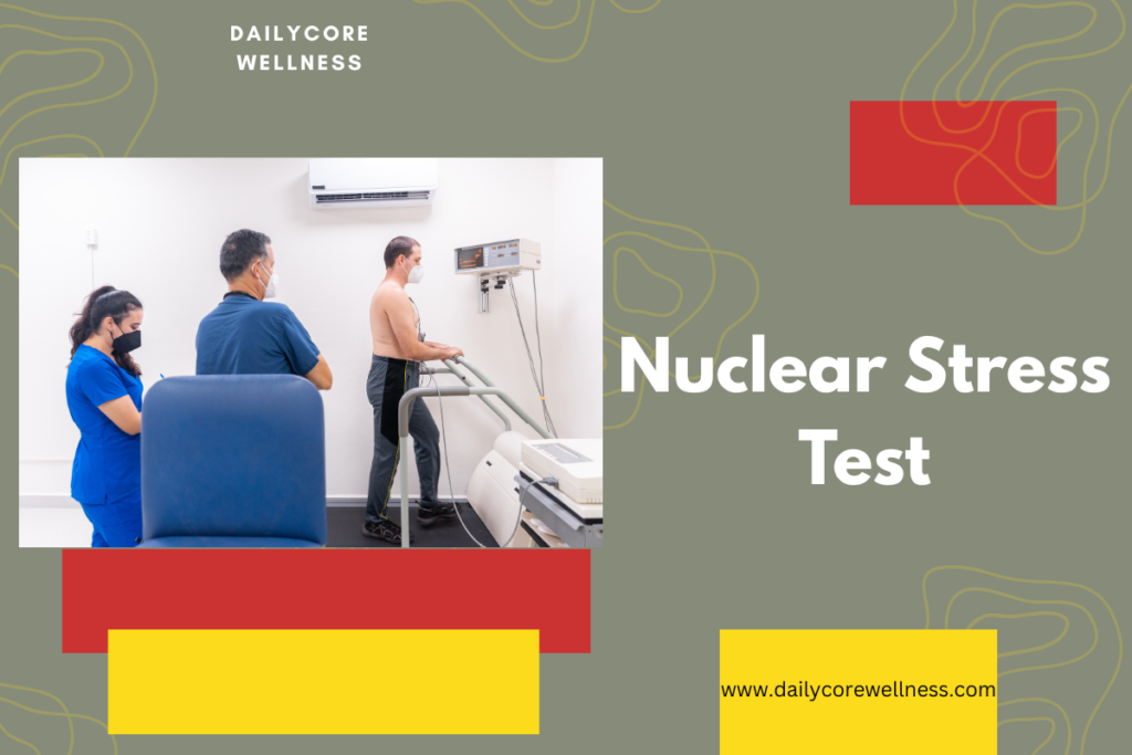
The Stress Phase: You will move to a treadmill, where ECG leads are attached to your chest to continuously monitor your heart rate and rhythm. The treadmill starts slowly and gradually increases in speed and incline every few minutes. Once your heart reaches its target rate, a second dose of the tracer is injected through your IV. You will walk for one additional minute to allow the tracer to circulate at peak exertion.
-
Note: If you are unable to exercise on a treadmill due to arthritis, mobility issues, or medical conditions, a pharmacological stress test is performed instead. Medications such as regadenoson, adenosine, or dobutamine are administered through your IV to simulate the effects of exercise by dilating your coronary arteries or increasing your heart rate.
-
-
Stress Imaging Phase: After the stress portion, you will rest again while your heart rate returns to normal. You will then lie under the camera a second time so the team can take stress images to compare directly against your resting baseline scans.
Understanding the Results
A board-certified cardiologist or nuclear radiologist compares the resting images with the stress images side-by-side to assess blood perfusion. The results typically fall into three primary categories:
| Result | Resting Images | Stress Images | What It Means |
| Normal | Normal blood flow | Normal blood flow | Blood flow to your heart is adequate both at rest and under stress. Coronary artery disease is unlikely. |
| Reversible Defect (Ischemia) | Normal blood flow | Reduced blood flow | Blood flow is normal at rest but restricted during stress. This indicates one or more severely narrowed coronary arteries that need treatment. |
| Fixed Defect (Scarring) | Reduced blood flow | Reduced blood flow | Blood flow is persistently reduced at both rest and stress. This typically indicates permanent heart muscle damage from a past heart attack. |
Safety and Risks
Nuclear stress tests are widely considered safe diagnostic procedures. The amount of radiation exposure from the tracer is low—roughly equivalent to the background radiation you naturally absorb from the environment over a few years—and the tracer passes naturally out of your body through urine within a day or two (drinking plenty of fluids after the test speeds up this process).
Minor side effects from the stress phase or pharmacological agents may include flushing, headache, dizziness, mild chest discomfort, or shortness of breath. Medical staff continuously monitor your blood pressure, ECG, and symptoms throughout the entire procedure to ensure immediate care if any complications arise.
What Happens After a Nuclear Stress Test?
Once both the resting and stress imaging phases are complete, your IV line will be removed, and you can generally return to your normal daily activities right away. Unless your doctor gives you specific instructions—such as holding certain cardiac medications longer or resting—you can resume driving, eating, and working as usual.
To help your body naturally flush out the radioactive tracer, you should drink plenty of water and fluids over the next 24 to 48 hours. The tracer has a short half-life and leaves your body primarily through urine and stool. While the radiation level is extremely low and poses minimal risk to people around you, standard guidance suggests washing your hands thoroughly after using the bathroom and avoiding close, prolonged contact with infants or pregnant women for the remainder of the day out of an abundance of caution.
Potential Follow-Up Actions Based on Your Results
The findings from your nuclear stress test provide your healthcare team with a clear roadmap for your cardiovascular care:
-
If Your Test Is Normal: If blood flow is uniform during both rest and exercise, your doctor can largely rule out significant coronary artery blockages as the source of your symptoms. Focus will then shift to managing general heart disease risk factors through lifestyle modifications, such as maintaining a healthy diet, exercising regularly, and managing cholesterol or blood pressure.
-
If Your Test Shows Reversible Ischemia: If reduced blood flow appears only during the stress phase, it indicates significant arterial narrowing. Your cardiologist may adjust your medications (such as adding anti-anginal drugs, statins, or blood thinners) or recommend a cardiac catheterization (coronary angiogram). An angiogram allows doctors to visualize the exact location of the blockages and potentially fix them using angioplasty or stent placement.
-
If Your Test Shows a Fixed Defect: Persistent areas of reduced blood flow indicate past heart muscle damage or scarring (often from a previous, sometimes unrecognized, heart attack). Your care plan will focus on protecting remaining healthy heart tissue, managing heart failure symptoms if present, and preventing future cardiac events through targeted medical therapy and cardiac rehabilitation.
When to Contact Your Doctor
Types of Radioactive Tracers Used
Not all nuclear stress tests use the same radiopharmaceutical tracer. Depending on your medical history, kidney function, and the facility’s equipment, your doctor will select one of three primary imaging agents:
-
Technetium-99m (Sestamibi or Tetrofosmin): The most common tracer used in modern nuclear cardiology. It emits clear gamma radiation that produces high-resolution 3D images with a single-photon emission computed tomography (SPECT) camera, while subjecting the body to a lower radiation dose than older agents.
-
Thallium-201: Historically the standard tracer, thallium behaves similarly to potassium in heart tissue. While less common today, it is still used in specific cases to evaluate viable heart tissue vs. permanent scarring because it redistributes through heart cells over time.
-
Rubidium-82 or Ammonia N-13: Used specifically for PET (Positron Emission Tomography) nuclear stress tests. PET scans offer higher diagnostic accuracy and lower radiation exposure than traditional SPECT scans, making them particularly useful for patients with higher body mass index (BMI) or complex vascular conditions.
Comparing SPECT vs. PET Nuclear Stress Tests
Nuclear cardiology relies on two distinct imaging technologies, each offering unique technical advantages:
| Feature | SPECT Nuclear Stress Test | PET Nuclear Stress Test |
| Primary Tracer Used | Technetium-99m, Thallium-201 | Rubidium-82, Ammonia N-13 |
| Total Test Duration | 2 to 4 hours | 30 to 60 minutes |
| Image Resolution | High clarity | Exceptional clarity (better for soft tissue/BMI) |
| Radiation Exposure | Slightly higher | Lower |
| Main Advantage | Widely available in most hospitals/clinics | Measures absolute quantitative blood flow in mL/min |
Limitations and False Results
While a nuclear stress test is highly accurate (with sensitivity and specificity around 85–90%), it is not infallible. Understanding its limitations helps explain why doctors sometimes order additional testing:
-
Balanced Three-Vessel Disease: In rare cases where all three major coronary arteries are equally and severely narrowed, the camera may show “uniform” blood flow because no single area looks significantly worse than another. This can cause a false-normal result.
-
Artifact Interference: Soft tissue—such as dense breast tissue in women or a high diaphragm in men—can occasionally block gamma rays from reaching the camera, creating an artificial “shadow” that mimics a blockage (a false positive).
-
Patient Movement: Lying completely still under the scanner is essential. Moving even slightly during the 15-minute imaging capture can blur the scans, requiring repeat acquisitions.
Long-Term Heart Health Management
A nuclear stress test provides a snapshot of your heart’s current blood supply, but protecting your cardiovascular system requires ongoing management. Regardless of whether your results show mild ischemia or clean arteries, key preventative steps include maintaining an optimal blood pressure below 120/80 mmHg, keeping LDL cholesterol within your target range, engaging in 150 minutes of moderate aerobic exercise weekly, and managing daily stress levels.
A Positron Emission Tomography (PET) nuclear stress test is an advanced cardiac imaging procedure that uses short-lived positron-emitting radiotracers to evaluate myocardial blood flow and cellular metabolism. Unlike conventional SPECT scans that provide relative comparisons of blood distribution, PET offers precise, high-resolution quantitative measurements.
How a PET Nuclear Stress Test Works
A cardiac PET stress test relies on advanced physics and rapid-decay radiopharmaceuticals to capture clear cross-sectional images of the heart muscle.
-
Radiotracer Administration: Short-lived radioactive tracers—most commonly Rubidium-82 or Ammonia N-13—are injected through an IV. These isotopes emit positrons as they decay.
-
Positron Annihilation & Signal Detection: When an emitted positron collides with an electron in heart tissue, they annihilate each other, releasing two high-energy gamma photons traveling in opposite directions (180 degrees apart). The ring detectors in the PET scanner detect these simultaneous photon pairs to pinpoint exact locations in the heart.
-
Pharmacological Stress: Because the radioactive tracers decay extremely fast (Rubidium-82 has a half-life of only 75 seconds), PET stress tests almost exclusively use IV pharmacological agents (such as regadenoson, adenosine, or dipyridamole) to dilate coronary arteries within seconds rather than treadmill exercise.
-
Image Acquisition: The scanner captures rest and stress images back-to-back, completing the entire scanning process in roughly 30 to 45 minutes with minimal radiation exposure to the patient.
Why Measuring Absolute Blood Flow Matters
Traditional SPECT nuclear tests measure relative blood flow—they compare the brightest (best-perfused) area of the heart muscle to darker areas. If one region receives 50% less blood than another, SPECT identifies a defect. However, this relative comparison creates critical diagnostic blind spots that PET resolves by quantifying Absolute Myocardial Blood Flow (MBF) in exact units (milliliters of blood per minute per gram of heart tissue, mL/min/g).
1. Detecting Balanced Three-Vessel Disease
When all three main coronary arteries are equally and severely narrowed, relative SPECT imaging can be fooled because blood flow decreases uniformly across the entire heart. To a SPECT camera, the scan looks “normal” because no single area is significantly darker than another. PET quantifies the exact flow value (e.g., finding that flow is globally depressed at 0.6 mL/min/g instead of the normal stress level above 2.0 mL/min/g), preventing false-negative diagnoses in high-risk patients.
2. Calculating Coronary Flow Reserve (CFR)
PET measures both resting blood flow and peak stress blood flow to calculate the Coronary Flow Reserve (CFR):
A normal CFR ratio is typically greater than 2.0. A reduced CFR indicates that the coronary arteries cannot dilate properly to meet increased metabolic demand, offering a sensitive marker for early-stage cardiovascular disease.
3. Uncovering Microvascular Dysfunction
Many patients—particularly women and individuals with diabetes—experience chest pain caused by Coronary Microvascular Disease (CMD), where the microscopic blood vessels supplying the heart are damaged even though the major epicardial arteries show no large blockages. Traditional angiograms and SPECT scans cannot see these tiny vessels. PET quantifies abnormal MBF and reduced CFR, diagnosing microvascular dysfunction that would otherwise go undetected.
4. Superior Image Quality in High-BMI Patients
PET scanners use high-energy photons and built-in CT attenuation correction, which minimizes image distortion caused by soft tissue, diaphragmatic shadows, or higher body mass index (BMI). This produces sharper images and drastically reduces false-positive results.
| Feature / Modality | SPECT Stress Test | PET Stress Test | Stress Cardiac MRI (CMR) |
| Primary Mechanism | Single-photon gamma radiation from injected isotopes | Positron annihilation gamma detection | Magnetic fields, radio waves, and gadolinium contrast |
| Ionizing Radiation | Moderate to High (~9–12 mSv) | Low to Moderate (~2–3 mSv) | Zero radiation |
| Blood Flow Measurement | Relative flow only | Absolute flow (mL/min/g) & Coronary Flow Reserve | Relative flow & Late Gadolinium Enhancement (LGE) |
| Spatial Resolution | Moderate (4–10 mm) | High (4–5 mm) | Highest (<1.5 mm) |
| Diagnostic Accuracy | ~80–85% | ~90–95% | ~90–95% |
| Test Duration | 2 to 4 hours | 30 to 45 minutes | 45 to 60 minutes |
| Availability | Widespread (Most hospitals) | Moderate / Growing | Limited (Specialized centers) |
SPECT Stress Test
Pros:
-
Ubiquitous Access: Available in virtually all community hospitals and outpatient cardiology clinics.
-
Treadmill Friendly: Easily combined with physical treadmill exercise rather than relying strictly on pharmacological drugs.
-
Extensive History: Decades of clinical trial data validate its risk stratification accuracy.
Cons:
-
Attenuated Imaging: Vulnerable to image artifacts caused by soft tissue, dense breast tissue, or high BMI.
-
Relative Flow Limitation: Can miss balanced three-vessel disease (triple-vessel CAD).
-
Higher Radiation: Delivers the highest radiation dose among the three modalities.
PET Stress Test
Pros:
-
Quantitative Precision: Measures absolute myocardial blood flow to detect microvascular disease and balanced triple-vessel blockages.
-
Fast & Low Radiation: Completed in under an hour with significantly lower radiation exposure than SPECT.
-
Ideal for High-BMI Patients: CT-based attenuation correction neutralizes soft tissue distortion.
Cons:
-
Cost & Equipment: Requires on-site or nearby cyclotrons or specialized generators (e.g., Rubidium-82), making it expensive.
-
Exercise Limitations: Extremely short tracer half-lives require pharmacological stress rather than treadmill exercise.
Stress Cardiac MRI (CMR)
Pros:
-
Zero Radiation: Completely safe regarding ionizing radiation, making it ideal for younger patients or repeat scans.
-
Gold Standard for Structure: Unmatched spatial resolution for evaluating heart muscle anatomy, scar tissue, tissue inflammation, and pericardial disease.
-
Scar Tissue Differentiation: Late Gadolinium Enhancement (LGE) precisely distinguishes transmural infarction (dead tissue) from viable muscle.
Cons:
-
Patient Constraints: Cannot be used on patients with non-MRI-compatible pacemakers/defibrillators, severe claustrophobia, or metallic implants.
-
Operator Dependent: Requires highly specialized technicians and dedicated software analysis.
-
Renal Risks: Gadolinium contrast carries risks for patients with severe end-stage kidney disease (GFR <30 mL/min).
Summary Verdict: Which Is Best?
-
Choose SPECT for routine, widely accessible testing when physical treadmill performance needs to be evaluated.
-
Choose PET for high-BMI individuals, suspected microvascular disease, or complex multi-vessel CAD.
-
Choose Cardiac MRI when zero radiation is preferred, or when detailed structural anatomy, tissue viability, and scar mapping are needed alongside perfusion.